| Key
Facts |
- Common medical
problem, related to smoking
- Centriacinar most
common, other types: panlobular, paraseptal, irregular
- Chest radiography
insensitive for mild disease
- Centriacinar
predominately involves upper lung zones
- Panlobular predominately
involves lower lung zones
|
| Imaging Findings |
|
Chest radiography
- Hyperinflation
- flat
diaphragms
- widened
retrosternal air space
- lung height
increased
- small narrow
heart
- Parenchymal areas
of hypoattenuation
- inhomogeneous
distribution
- arterial
deficiency, increased branching angle of remaining vessels
- bullae
- increased
marking not clearly understood, combination of bronchial wall
thickening or superimposition of emphysematous
walls
- Secondary
manifestations
- Pulmonary artery
hypertension
- enlarged
central pulmonary arteries and peripheral arterial
pruning
- Sensitivity poor
for early disease, rare false positives
- problem is
recognition of loss of normal lung
- normal lung at
chest radiography is 90% air, making detection of slight increases in
air nearly impossible
- Crude correlation
between indices of airway obstruction and radiographic findings
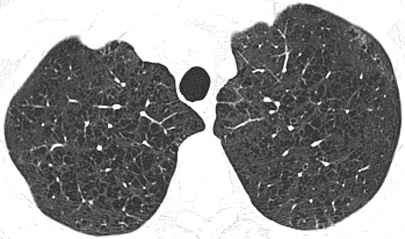
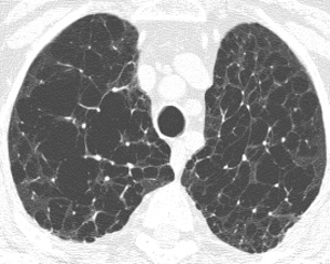
CT/HRCT
- More sensitive than
chest radiography
- Emphysematous holes
usually have no discernable wall
- Central artery may
remain visible surrounded by destroyed lung
- Objectively
measured by assuming that lung with a threshold HU < -960 is
emphysematous lung
|
| Differential Diagnosis |
- Technical: false
negatives with low dose technique or wide windows
- Asthma
- Contrictive
Bronchiolitis Obliterans
- Athletic hyperinflation
|
| Pathological Features |
- Abnormal
enlargement of the airspaces distal to the terminal bronchioles
accompanied by destructive changes of the alveolar walls without obvious
fibrosis
- Emphysema usually
inhomogeneous
- centriacinar
emphysema strongly associated with cigarette smoking
- Centriacinar:
dilatation 2nd order respiratory bronchioles in secondary
lobule
- Panlobular: involves
entire lobule
|
| Clinical Presentation |
- Dyspnea, shortness
of breath
- Obstructive pulmonary
function tests
- Treatment
- smoking cessation
- Bronchodilators
- Pneumococcus
vaccination
- Lung volume reduction
surgery (criteria, inhomogeneous distribution upper lung zones
- Lung Transplant
|
| References |
|
Stern EJ et al: CT of
the lung in patients with pulmonary emphysema: Diagnosis, quantification,
and correlation with pathologic and physiologic findings. AJR 162: 791-8,
1994.
|