| Key
Facts |
- Common
systemic disease
- Etiology
unknown
- Widespread
noncaseating granulomas, that resolve or cause fibrosis
- May
have associated erythema nodosum, uveitis, hypercalcemia, arthritis
- 95%
of patients have abnormal chest radiograph with lymphadenopathy and
or lung opacities
- HRCT
centriacinar nodules along bronchovascular bundles, septa,and periphery
of lobule
- Definitive
diagnosis with transbronchial, lymph node, or liver biopsy
- Most
patients have a good prognosis with resolution of findings in < 2
years
- Major
complications include respiratory failure from fibrosis, mycetomas,hemorrhage,
and cor pulmonale
|
| Imaging
Findings |
| Chest
radiography
- Abnormal chest
radiograph – 95%
- 5 to 15% have
normal chest radiograph at onset
- Lymphadenopathy,
(80%) most common finding – bilateral hilar/paratracheal
Usually not visible at 2 years; may persist for many years
Nodes may calcify, sometimes eggshell calcification
- Lung disease,
(<50%), often manifest with nodal regression
- Pattern
and Distribution:
- Reticulonodular
opacities (90%)
- Large airspace
nodules with air bronchograms
- Fibrosis
mid and upper lung zones
- Upper lobe
cyst formation with aspergilloma
Stage 0 –
clear chest radiograph (5 – 15%, at presentation)
Stage 1 –
lymphadenopathy (45 – 65%); 60% resolve completely
Stage 2 –
lymphadenopathy and lung opacities (30 – 40%)
Stage 3 –
Lung opacities (10 – 15%)
Stage 4 –
fibrosis with or without lymphadenopathy (5 – 25%)
- Atypical
appearance
- Atypical
lymphadenopathy - unilateral hilar, posterior mediastinal
- Airway
compression
- Unilateral
lung disease
- Cavitary
lung lesions
- Pleural
effusion
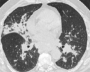
CT/HRCT
- CT may show lymphadenopathy
at left paratracheal, AP window, anterior mediastinum, retroperitoneal
- HRCT, may be abnormal
with normal CXR
- Predominantly involves
the mid and upper lung zones
- Pattern: 1 –
5 mm centrilobular nodules along brochovascular structures, septa, and
periphery of lobule
- Often extends in
a swath from the hilum to lung periphery
- Ground glass opacities
- Progressive massive
fibrosis, distortion, honeycombing, cysts, bullae, traction bronchiectasis
- Mycetomas in cavities
and cysts
- Large and small
airway stenoses
|
| Differential
Diagnosis |
- Granulomatous
diseases : TB, fungal infection, berylliosis, extrinsic allergic alveolitis
- Diseases with granuloma-like
reactions: lymphoma, carcinoma, metastases
- Chronic eosinophilic
pneumonia, BOOP
|
| Pathological
Features |
- Common systemic
disease
- Widespread noncaseating
granulomas, that resolve or cause fibrosis
- Etiology unknown
- Well-formed granulomas
; central epitheliod histiocytes and multinucleate giant cells surrounded
by lymphocytes, monocytes and fibroblasts
- Lymphatic distribution
|
| Clinical
Presentation |
- 10 times more
common in blacks, female predominance
- Onset – usually
age 20 to 40
- Asymptomatic, or
fatigue, malaise, weight loss, fever, respiratory symptoms, erythema
nodosum, uveitis, skin lesions, arthropathy, bone lesions
- In < 2% TB precedes
sarcoidosis or develops later
- 80% of cases resolve
completely; in 20% fibrosis develops that may destroy and distort the
lung
- Anemia, leukopenia,
elevated sedimentation rate, hypercalcemia, nephrolitiasis
- Cutaneous anergy
- Raised ACE level,
not specific
- Diagnosis from
lung, lymph nodes, and liver
- Bronchoscopy and
transbronchial biopsy – positive in 90% of cases, even if CXR
is normal
- BAL – increased
CD4/CD8 ratio, nonspecific finding
- Usually not treated;
steroids in severe cases
- Recurrence in transplanted
lung has been reported
- Prognosis worse
in blacks
- Mortality – 2 – 7%; death from respiratory
failure, cor pulmonale, hemorrhage
|
| References |
Miller
BH, Rosado-de-Christenson ML, McAdams HP, et al. Thoracic sarcoidosis:
radiologic-pathologic correlation Radiographics 15:421-437, 1995
Rockoff SD , Rohatgi PK. Unusual manifestations of thoracic sarcoidosis
AJR 144:513-528, 1985
Traill ZC, Maskell GF , Gleeson FV. High-resolution CT findings of pulmonary
sarcoidosis AJR 168:1557-1560, 1997
|