| Key
Facts |
- Permeation of lymphatics
by neoplastic cells
- Tumor emboli or
direct spread to lungs from hilar nodes or lung cancer mass
- Seen with carcinoma
of the lung, breast, pancreas, stomach, colon, prostate and other tumors
- Unilateral disease
- most commonly due to lung cancer
- Radiography - may
resemble interstitial edema; progressive disease
- HRCT: nodular thickening
of interlobular septa and bronchovascular bundles
- Lung architecture
preserved
- Prognosis - poor
|
| Imaging
Findings |
Chest
radiography
- Reticulonodular
opacities, coarse bronchvascular markings, septal lines, subpleural
edema at fissures
- May resemble interstitial
edema
- Hilar and mediastinal
lymphadenopathy may be present
- Pleural effusion,
common
- Unilateral disease
- most commonly due to lung cancer
- Bilateral symmetric
disease commonly due to extrathoracic primary tumor
- Chest radiograph
may be normal
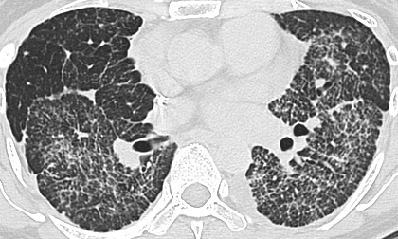
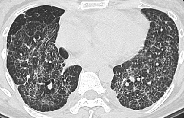
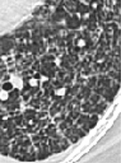
CT/HRCT
- HRCT best imaging
to suggest diagnosis
- Nodular thickening
of interlobular septa and bronchovascular bundles
- Septal lines and
polygons with nodular or beaded appearance
- Lung architecture
preserved
- Patchy ground glass
and airspace opacities
- Small centrilobular
nodules, thickened centrilobular bronchovascular bundles
- Peripheral or central
distribution, basal predominance
- Commonly asymmetric,
may spare lobes or lungs
- Smooth or nodular
thickening of interlobar fissures
- Pleural effusion
- Hilar/mediastinal
lymphadenopathy
|
| Differential
Diagnosis |
- Pulmonary edema
- Interstitial pneumonia
- UIP
- Scleroderma
- Drug reaction
- Sarcoid
- Asbestosis
- Hypersensitivity
pneumonitis
- Differentiation
- Lymphangitic
carcinomatosis will not show architectural distortion or honeycombing.
Progressive disease. Usually not occult but develops in patients with
known malignancy
- Progression of
disease without treatment
|
| Pathological
Features |
- Frequent form of
tumor spread
- Permeation of lymphatics
by neoplastic cells
- Interstitial thickening
of interlobular septa due to tumor cells, desmoplastic response, and
dilated lymphatics
- Hilar and mediastinal
lymph nodes may or may not be involved
- Pathogenesis
- Hematogenous
metastases i.e., tumor emboli to small pulmonary artery branches with
subsequent spread along lymphatics
- Some tumors such
as lymphoma spread from hilar nodes retrograde into pulmonary lymphatics
- Lung cancer can
spread to adjacent lung along lymphatics
|
| Clinical
Presentation |
- Any age, male or
female
- Seen with carcinoma
of the lung, breast, pancreas, stomach, colon, prostate and other tumors
- Dyspnea, cough,
progressive symptoms
- Diagnosis evident
in patients with known malignancy
- If no known malignancy
– sputum cytology, transbronchial biopsy, fine needle aspiration
biopsy or open lung biopsy for diagnosis
- Poor prognosis,
15% survive 6 months
|
| References |
Trapnell
DH. Radiological appearance of lymphangitis carcinomatosa of the lung
Thorax 19:251-260, 1964
Ren H, Hruban RH, Kuhlman JE, et al. Computed tomography of inflation-fixed
lungs: the beaded septum sign of pulmonary metastases J Comput Assist
Tomogr 13:411-416, 1989
|