Radiographic interpretation requires a firm knowledge of normal anatomy and the myriad variants that may simulate disease.
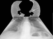
| Cross-sectional Anatomy Learn basics of cross-sectional anatomy with either anatomic sections (from NIH visible human) or from radiographic sections (normal computed tomography scan). Seventy-four anatomic structures for you to identify. Ten cross-sectional planes (10 cadaveric, 10 mediastinal windows and 10 lung windows) (total download 828K) |
 |
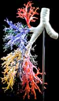
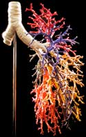
| Right Bronchial Anatomy | Left Bronchial Anatomy High detail 3D anatomy of the bronchial airways. Rotate the airways 360 degrees. Zoom in on the detail of any airway. Each segmental airway is color coded. (total file size 16 Mb, requires Quicktime) |
  |
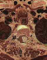
| Mediastinal Anatomy Detailed cross-sectional anatomy illustrated with anatomic sections from NIH visible human. Twenty-three cadaveric sections – 57 anatomic structures labelled. (total download 984K) |
 |
|
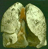
Inflated Lungs – Virtual Reality from the University of Arkansas Department of Anatomy Note the curvilinear course of the fissures. Black anthracotic pigment outlines numerous secondary pulmonary lobules, especially along the dorsal aspect of the lung. The edge of the lung along the right middle lobe and lingula is thin, which makes these areas a favorite site for open surgical biopsy. |
 |
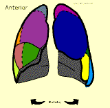
| Segmental Projections Rotate the lungs, each segment is color coded and labelled. Projections similar to perfusion scanning in nuclear medicine. (total download 55K) |
 |
| Introduction to Cardiothoracic Imaging C. Carl Jaffe, MD and Patrick J. Lynch, MS Yale University Anatomy, Techniques including Echocardiography, Radiographic Findings, and multiple cases studies |
|